
How ear wax forms in the ear canal
- 1 day ago
- 10 min read
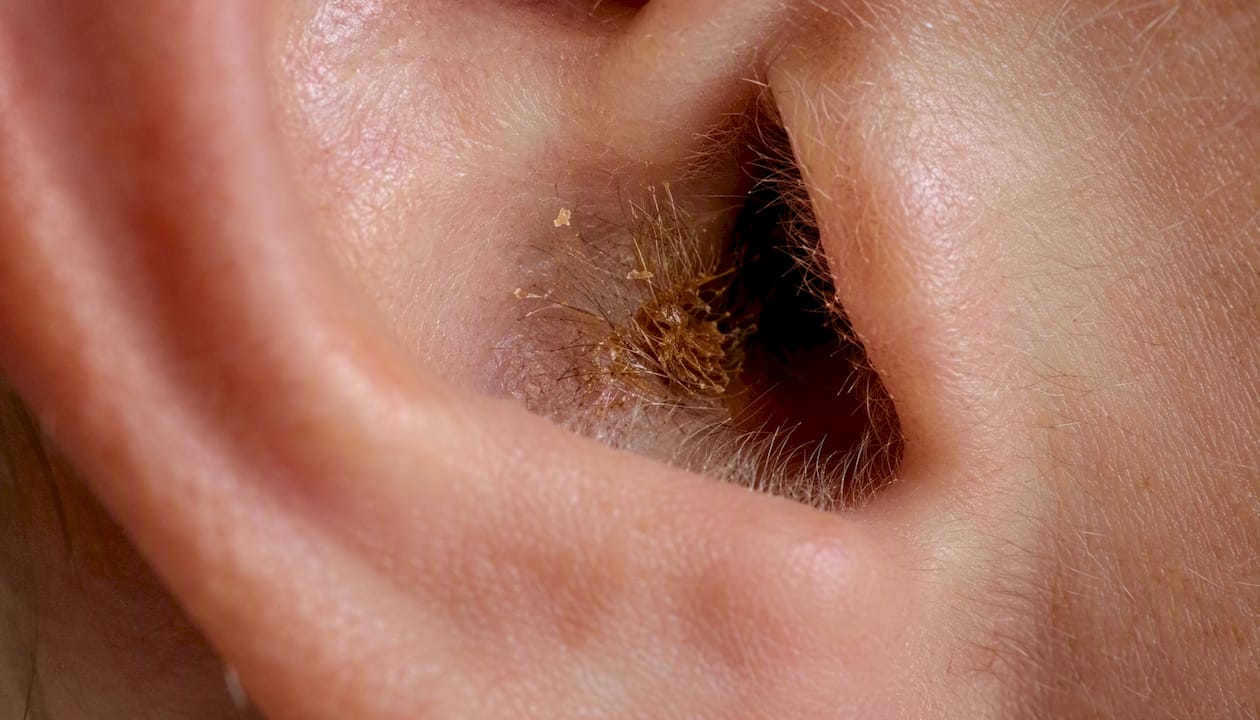
Ear wax, known clinically as cerumen, is produced by two specialised glands in the outer one-third of the ear canal: the ceruminous glands and the sebaceous glands. Their secretions mix with shed skin cells, fine hairs, and environmental debris to create the waxy substance you may notice at the entrance to your ear. Far from being a sign of poor hygiene, cerumen protects and lubricates the ear canal, trapping dust and microbes before they can reach the delicate eardrum. Under normal circumstances, the ear clears itself gradually through a process called epithelial migration, assisted by the everyday movement of your jaw. Most wax dries, flakes, and falls away unnoticed.
Key takeaways
Ear wax forms when ceruminous and sebaceous glands in the outer ear canal produce secretions that mix with shed skin and debris, and most wax clears naturally without any intervention.
Point | Details |
Where wax forms | Ceruminous and sebaceous glands in the outer, cartilaginous one-third of the ear canal. |
Normal clearance | Epithelial migration and jaw movement carry wax outward; most falls away as dry flakes unnoticed. |
When it becomes a problem | Impaction causes hearing loss, fullness, pain, tinnitus, or itchiness and warrants professional assessment. |
Safe home measures | Olive oil, sodium bicarbonate, or cerumenolytic drops can soften mild build-up; never insert objects into the canal. |
When to book professional care | Persistent symptoms, sudden hearing loss, or pain require removal by a trained, HIS- or CQC-regulated clinician. |
Where in the ear canal is wax actually produced?
The ear canal is roughly 2.5 cm long and divides into two distinct zones. The outer, cartilaginous third is where all cerumen production takes place. The inner, bony section contains no glands at all and is covered by thin, tightly adherent skin that cannot tolerate the same kind of manipulation.
Two types of exocrine gland sit within the skin of the outer canal, both closely associated with hair follicles:
Ceruminous glands: Modified apocrine sweat glands that secrete a watery, protein-rich fluid. They are the primary source of the characteristic waxy component of cerumen.
Sebaceous glands: Oil-secreting glands that contribute sebum, giving cerumen its lubricating, water-repellent properties.
These ceruminous and sebaceous glands sit alongside the fine hairs of the outer canal. When their secretions combine with shed keratinocytes (dead skin cells) and any debris that has entered the canal, the result is cerumen. The bony inner canal lacks this glandular architecture entirely, which is one reason pushing wax inward with a cotton bud or finger is so counterproductive: you are moving material away from its natural exit point and into a zone with no self-cleaning mechanism.
Pro Tip: A simple labelled diagram of the ear canal, showing the cartilaginous outer third, the two gland types, and the bony inner section, is an excellent visual aid to place here. Readers who can see the anatomy grasp the self-cleaning logic far more readily than those who rely on text alone.

For more on ear anatomy and common conditions, the ear facts resource at Earhealthservice covers these structures in accessible detail.
What is ear wax made of, and why does it look different in different people?
Cerumen is not a single substance. It is a mixture of several components, each contributing to its protective and lubricating functions.
Major components:
Sebum from sebaceous glands
Ceruminous gland secretions (water, proteins, lipids)
Keratin from shed skin cells
Fine canal hairs
Dust, microbes, and environmental debris
The lipid fraction of cerumen has been analysed in some detail. Squalene accounts for a notable fraction of cerumen’s composition, and cholesterol is also a significant component, while shed skin (keratin) forms a substantial portion of the overall mass.
Component | Role in cerumen |
Squalene (12–20%) | Antimicrobial lipid; contributes to water resistance |
Cholesterol (6–9%) | Structural lipid; part of the sebaceous fraction |
Keratin (shed skin) | Substantial bulk of the wax mass |
Proteins and fatty acids | Antimicrobial and lubricating properties |
Hair and debris | Trapped particles carried outward |
Wet versus dry cerumen: a genetic difference
Cerumen comes in two broad types: wet (honey-brown, soft) and dry (grey, flaky). The difference is almost entirely genetic, determined by a single nucleotide polymorphism in the ABCC11 gene. Wet cerumen is the dominant trait and is more common in people of European and African ancestry. Dry cerumen is more prevalent in East Asian and some Indigenous populations. Neither type is healthier than the other, though dry cerumen can sometimes be more prone to compaction in certain canal shapes.
How does ear wax normally move out of the ear?
The ear is one of the few places in the body where skin migrates outward rather than shedding in place. This process, called epithelial migration, begins at the eardrum and moves slowly toward the canal entrance, carrying cerumen with it. Jaw movement during chewing and talking accelerates this outward flow, which is why the ear canal is remarkably good at self-maintenance in most people.
The main forces that move cerumen outward are:
Epithelial migration: Skin cells at the eardrum divide and move laterally toward the canal opening at roughly the same rate as a fingernail grows.
Hair shaft transport: Fine hairs in the outer canal act as a physical guide, directing wax toward the entrance.
Jaw and mastication movement: Chewing creates vibration and slight canal-wall movement that dislodges and advances cerumen.
Mild desiccation: As wax reaches the outer canal, it dries slightly and breaks into flakes that fall away naturally.
Inserting objects into the ear canal disrupts this outward flow. A cotton bud, for instance, may remove a small amount of surface wax but simultaneously compacts the deeper mass against the bony canal wall, where no glands or migration mechanism can shift it. This is one of the most common reasons people develop impaction despite regular cleaning attempts.
What causes ear wax buildup in some people more than others?
Production and consistency are shaped primarily by genetics, age, gland density, and skin type. Some people simply produce more cerumen than others, and that is entirely normal. The problems arise when production outpaces clearance, or when clearance is disrupted.
Common factors that influence cerumen production and build-up:
Genetics: Wet cerumen types tend to be stickier and accumulate more readily than dry types.
Age: Cerumen becomes drier and less mobile with age, and the jaw movement that aids clearance may reduce. Older adults are disproportionately affected by impaction.
Narrow or hairy canals: A narrower canal or dense hair growth slows the outward migration of wax considerably.
Hearing aids and earbuds: Devices that sit in the canal block the natural exit route and can stimulate additional secretion through mild irritation.
Skin conditions: Eczema, psoriasis, and seborrhoeic dermatitis alter the skin’s shedding pattern and can change cerumen consistency.
Dusty or humid environments: Higher debris load or moisture can alter the texture of cerumen and increase the rate of accumulation.
Medications and hormonal changes: Some medications affect sebaceous gland activity; hormonal shifts can alter lipid secretion.
Repeated cleaning with cotton buds creates a particular problem. Rather than removing wax, it tends to compact it and can abrade the delicate canal skin, triggering increased gland secretion as a protective response. People who repeatedly attempt self-cleaning often present with harder, deeper impactions than those who leave their ears alone.
Pro Tip: Patients with narrow canals, prominent hair growth, or high sebaceous activity are the most likely to develop hard impactions. If you find yourself needing to clean your ears frequently, that pattern itself is a signal worth discussing with a clinician rather than managing with cotton buds.
When does ear wax become a problem?
Cerumen becomes clinically significant when it accumulates to the point of blocking the canal, a condition called cerumen impaction. Common symptoms include hearing loss, a sensation of ear fullness, pain, tinnitus, itchiness, and discharge. The hearing loss is conductive: sound waves cannot travel efficiently through a blocked canal to reach the eardrum.
Symptoms that suggest impaction:
Muffled or reduced hearing (conductive hearing loss)
A feeling of pressure or fullness in the ear
Earache or discomfort
Tinnitus (ringing, buzzing, or hissing sounds)
Itchiness inside the canal
Discharge or an unpleasant odour
Seek a routine appointment with a trained clinician if you notice any of the above symptoms persisting for more than a few days. Seek more urgent care if you experience sudden hearing loss, severe pain, vertigo, fever, or any discharge that looks like blood or pus. These may indicate a perforated eardrum or infection, and self-treatment would be inappropriate.
Per current NHS guidance, microsuction is the preferred removal method in many UK clinical pathways. Trained Aural Care Specialists assess each patient individually and select the safest procedure based on medical history and the clinical presentation of the ear. For guidance on when to seek urgent care, Earhealthservice’s urgent ear wax treatment workflow sets out the referral pathway clearly.
Safe home measures and professional removal options
For mild, uncomplicated wax build-up, softening the wax first is a reasonable starting point. The goal is to make the cerumen less viscous so the ear’s natural clearance mechanism can do its job.
Safe home options (aligned with NHS guidance):
Olive oil or almond oil drops: A few drops of warm (body temperature) oil in the affected ear, two or three times daily for up to two weeks, can soften wax sufficiently for it to migrate out naturally. Do not use if you have a known perforated eardrum or active ear infection.
Sodium bicarbonate drops: Available from pharmacies; effective at softening harder wax. Again, avoid with a perforated eardrum.
Proprietary cerumenolytic drops: Pharmacy products such as Earol or Otex are formulated for this purpose and carry clear instructions.
NHS inform advises that most earwax falls out naturally and that inserting objects into the ear canal, including cotton buds, should be avoided. If drops have not resolved the problem after two weeks, or if symptoms are significant, professional removal is the appropriate next step. For a fuller overview of home remedies, Earhealthservice’s guide to ear wax remedies covers the options in detail.
Professional removal methods
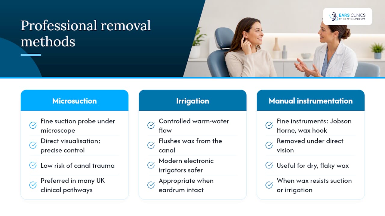
Three clinically validated techniques are used by trained Aural Care Specialists:
Microsuction uses a fine suction probe under direct visualisation with a microscope or loupe. Because the clinician can see the canal throughout, it is precise and carries a low risk of canal trauma. Microsuction’s direct visual control and minimal canal irrigation make it the preferred technique in many UK clinical pathways and training programmes. It is suitable for most patients, including those with perforated eardrums or a history of ear surgery.
Irrigation (previously called syringing) uses a controlled flow of warm water to flush wax from the canal. Modern electronic irrigators are considerably safer than older metal syringes. Irrigation is appropriate when the eardrum is intact and there is no history of perforation, ear surgery, or active infection.
Manual instrumentation uses fine instruments such as a Jobson Horne probe or wax hook to remove wax under direct vision. It is particularly useful for dry, flaky wax that does not respond well to suction or irrigation.
Pro Tip: If you wear hearing aids, schedule a clinic check every six to twelve months even if you have no symptoms. Hearing aids accelerate wax accumulation and can mask the early signs of impaction.
Ear care should only be performed by trained, competent clinicians following best practice standards and regulated by Healthcare Improvement Scotland (HIS) or Care Quality Commission (CQC). Attempting removal at home with candles, syringes, or improvised tools carries a genuine risk of eardrum perforation and canal injury.
What should you expect from a regulated UK ear clinic?
Regulated clinics staffed by trained Aural Care Specialists follow NICE and NHS safety standards throughout every appointment. The process is straightforward and, for most patients, entirely comfortable.
What a typical clinic visit involves:
A brief clinical history, including any previous ear surgery, perforations, or infections
Visual inspection of the ear canal and eardrum, often with before-and-after imaging
Informed consent and a clear explanation of the chosen removal method
The procedure itself, which usually takes 15–30 minutes
Aftercare advice and guidance on prevention
Earhealthservice operates EARS Clinics in Glasgow and Edinburgh, registered with Healthcare Improvement Scotland (HIS) and licensed to treat patients from two years of age. The clinics offer microsuction, irrigation, and manual instrumentation, with same-day appointments and home visits available where clinically suitable. Appointments for adults are priced at £60, with under-18s at £75 and home visits at £180. You can book directly at Earhealthservice.
For children attending loud events, appropriate outer ear noise protection is a sensible precaution alongside good ear hygiene.
Practical steps to reduce troublesome wax build-up
Simple, consistent habits reduce the likelihood of impaction without interfering with the ear’s natural self-cleaning process.
Do:
Clean only the outer ear (the visible part of the pinna) with a soft cloth.
Use softening drops preventively if you have a history of build-up, after discussing this with a clinician.
Keep hearing aids and earbuds clean and dry; remove them for periods each day to allow the canal to ventilate.
Schedule regular clinic checks if you wear hearing aids or have narrow canals.
Do not:
Insert cotton buds, fingers, ear candles, or any other object into the ear canal.
Use ear candles: there is no clinical evidence they are effective, and they carry a burn risk.
Attempt to irrigate your own ear at home with a syringe unless specifically instructed by a clinician.
Pro Tip: Hearing aid users benefit most from a scheduled six-monthly check rather than waiting for symptoms. By the time a hearing aid user notices reduced sound quality, the canal is often already significantly blocked.
For a detailed, practical prevention plan, Earhealthservice’s guide to preventing earwax buildup covers daily habits and clinic scheduling in full.
A clinician’s perspective on ear wax concerns
We see patients every week who have been managing wax build-up with cotton buds for years, convinced they are keeping their ears clean. In practice, they are often the patients who arrive with the most compacted wax and the most significant hearing loss. The ear’s self-cleaning mechanism is genuinely effective for most people most of the time, and the single most helpful thing you can do is stop interfering with it.
When wax does need removing, microsuction is our preferred approach because it gives the clinician direct visual control throughout. That said, the right method depends on your individual anatomy, your medical history, and the nature of the wax itself. Irrigation and manual instrumentation are equally valid when the clinical picture calls for them. What matters is that the decision is made by a trained, regulated clinician who has examined your ear, not by a general assumption about which technique sounds most advanced.
If you are concerned about your hearing or have noticed any of the symptoms described in this article, please do not wait. A single appointment with a qualified Aural Care Specialist is usually all it takes to resolve the problem safely and completely.
Sources
This article is general information, not a substitute for advice from a qualified doctor. Consult a qualified healthcare professional about your own circumstances before acting on anything here.
Recommended