
What is the tympanic membrane? Anatomy and function

TL;DR:
The tympanic membrane, or eardrum, is a layered, cone-shaped structure that converts sound waves into vibrations, enabling hearing. It also acts as a protective barrier and is supported by various muscles, with its health crucial for long-term auditory function. Damage or disease affecting the membrane can impair hearing and requires professional assessment to ensure proper healing and protection.
The tympanic membrane, commonly known as the eardrum, is defined as the thin, cone-shaped membrane separating the outer ear canal from the middle ear, converting airborne sound waves into mechanical vibrations that make hearing possible. It serves simultaneously as a sensory interface and a protective barrier, shielding the delicate middle ear structures from infection, debris, and pressure fluctuations. Understanding how this small but remarkable structure works helps you recognise when something is wrong and why professional ear care matters so much for long-term hearing health.
What is the tympanic membrane’s anatomy and structure?
The tympanic membrane is composed of three distinct microscopic layers: a lateral cutaneous (skin) layer facing the ear canal, an intermediate fibrous layer that provides structural rigidity, and a medial mucous layer facing the middle ear. Each layer contributes to the membrane’s ability to respond precisely to sound pressure while remaining durable enough to withstand daily exposure to the environment. This layered construction is what makes the eardrum both sensitive and resilient.

In terms of shape and orientation, the membrane is cone-shaped at approximately 55 degrees relative to the floor of the ear canal. This oblique angle is not incidental. It optimises the surface area exposed to incoming sound waves, improving the efficiency of vibration capture. The membrane is anchored around its circumference by a fibrocartilaginous ring that holds it firmly within the bony ear canal.
The membrane measures approximately 9 to 10 mm in its longest diameter and 8 to 9 mm in its shortest, making it roughly the size of a small fingernail. At its centre sits the umbo, the most depressed point of the membrane, which connects directly to the handle of the malleus bone. The umbo acts as the primary transfer point for vibrations moving from the membrane into the ossicular chain.
Anatomical feature | Description |
Layers | Cutaneous, fibrous, and mucous |
Shape and angle | Cone-shaped, oblique at approximately 55 degrees |
Dimensions | 9 to 10 mm by 8 to 9 mm |
Central landmark | Umbo, connected to the malleus |
Regions | Pars tensa (stiff, large) and pars flaccida (thin, flexible) |
The membrane is divided into two functional regions. The pars tensa and pars flaccida differ in thickness and stiffness, which means they respond differently to sound pressure. The pars tensa, which forms the majority of the membrane, is taut and responsible for most sound transmission. The pars flaccida, located in the upper portion, is thinner and more pliable, making it more susceptible to pathological changes such as retraction pockets.
Pro Tip: If you are ever shown an image of your eardrum during a clinical examination, the light reflex, a triangular bright spot visible in the lower front quadrant, confirms the membrane’s normal cone shape and healthy orientation.
How does the tympanic membrane work in sound transmission?
Sound transmission through the tympanic membrane follows a precise mechanical sequence. When sound waves travel down the ear canal and strike the membrane, it vibrates in response. These vibrations are then transferred to the three ossicles of the middle ear: the malleus, incus, and stapes, in that order.
Sound waves enter the ear canal and create pressure fluctuations against the tympanic membrane.
The membrane vibrates, with the umbo transmitting movement directly to the malleus handle.
The malleus transfers vibration to the incus, which passes it to the stapes.
The stapes footplate pushes against the oval window of the cochlea, converting mechanical vibration into fluid movement.
Fluid movement within the cochlea stimulates hair cells, which generate nerve signals interpreted by the brain as sound.
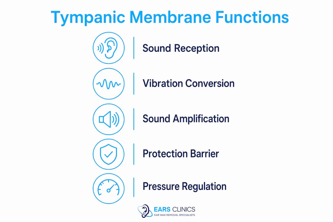
The conversion of airborne sound waves into mechanical vibrations is the membrane’s primary function, and the efficiency of this process directly determines hearing acuity. Any disruption to the membrane’s surface area, tension, or integrity reduces the fidelity of this conversion.
One aspect of tympanic membrane function that is frequently underappreciated is its role in sound amplification. Because the membrane’s surface area is considerably larger than the stapes footplate, the pressure of incoming sound is concentrated as it passes through the ossicular chain. This mechanical advantage allows the auditory system to detect even faint sounds with remarkable sensitivity. Without this amplification, the threshold for audible sound would be significantly higher, meaning quieter sounds would simply go unheard.
The relationship between the tympanic membrane and the cochlea is therefore not passive. The membrane actively shapes the quality and intensity of the signal before it ever reaches the inner ear, which is why even partial damage to the eardrum can produce disproportionate effects on hearing clarity.
What protective mechanisms support the tympanic membrane?
The tympanic membrane does not operate in isolation. Several biological safeguards work alongside it to protect both the membrane itself and the broader auditory system.
Tensor tympani and stapedius muscles: These two small muscles attach to the malleus and stapes respectively. When exposed to sudden loud sounds, they reflexively dampen ossicle movement, reducing the intensity of vibration transmitted to the inner ear. This acoustic reflex is an automatic protective response to acoustic trauma.
Eustachian tube: This narrow channel connects the middle ear to the back of the throat. Its primary role is to equalise air pressure on both sides of the tympanic membrane, allowing it to vibrate freely. When the Eustachian tube is blocked, such as during a cold or at altitude, the resulting pressure imbalance stiffens the membrane and reduces hearing acuity noticeably.
Barrier function: The membrane physically prevents bacteria, water, and foreign material from entering the middle ear. A healthy, intact eardrum is one of the body’s most effective defences against middle ear infection.
Self-repair capacity: Unlike many tissues, the tympanic membrane has a limited but genuine ability to heal small perforations spontaneously, provided the surrounding tissue remains healthy and infection-free.
Pressure imbalances, whether caused by flying, diving, or upper respiratory infections, can impair membrane vibration and temporarily reduce hearing. This is why ear health matters beyond simply avoiding loud noise. Managing conditions that affect the Eustachian tube is as important as protecting the membrane from direct trauma.
Pro Tip: Swallowing, yawning, or performing a gentle Valsalva manoeuvre (pinching the nose and blowing gently) can help open the Eustachian tube and relieve pressure discomfort during altitude changes. Never attempt this if you have an active ear infection.
What conditions affect the tympanic membrane and hearing?
Several clinical conditions can compromise tympanic membrane integrity and, by extension, hearing function. Recognising the signs early makes a significant difference to outcomes.
Common conditions include:
Tympanic membrane perforation: A hole or tear in the membrane caused by infection, trauma, a sudden pressure change, or a foreign object. Perforations impair sound transmission and leave the middle ear vulnerable to infection. Small perforations often heal without intervention, while larger ones may require surgical repair (myringoplasty).
Otitis media: Middle ear infection, frequently associated with fluid accumulation behind the membrane. The membrane may appear red, bulging, or opaque during examination. Repeated episodes can cause scarring and long-term hearing changes.
Tympanosclerosis: Calcium deposits that form within the membrane following repeated inflammation. These deposits stiffen the membrane and reduce its ability to vibrate efficiently, producing a degree of conductive hearing loss.
Retraction pockets: Areas where the pars flaccida is drawn inward due to chronic negative pressure, often linked to Eustachian tube dysfunction. Left untreated, retraction pockets can develop into cholesteatoma, a destructive growth requiring surgical management.
Condition | Primary cause | Hearing impact |
Perforation | Trauma, infection, pressure | Conductive hearing loss, infection risk |
Otitis media | Bacterial or viral infection | Temporary or persistent hearing reduction |
Tympanosclerosis | Repeated inflammation | Stiffened membrane, reduced vibration |
Retraction pocket | Eustachian tube dysfunction | Progressive hearing loss if untreated |
Symptoms that warrant prompt professional assessment include persistent ear pain, a feeling of fullness in the ear, sudden hearing loss, discharge from the ear canal, or ringing in the ear (tinnitus). Early treatment for ear conditions consistently produces better outcomes than delayed intervention. Maintaining membrane integrity through safe ear care practices and timely clinical review is the most reliable way to preserve hearing over the long term.
Key takeaways
The tympanic membrane is both a precision sound transducer and a protective barrier, and its integrity is the single most important factor in maintaining healthy hearing.
Point | Details |
Three-layer structure | The cutaneous, fibrous, and mucous layers give the membrane both sensitivity and durability. |
Sound amplification role | The membrane’s surface area advantage over the stapes footplate concentrates sound pressure for clearer hearing. |
Pressure regulation | The Eustachian tube must equalise pressure on both sides of the membrane for normal vibration to occur. |
Clinical vulnerability | Perforations, infections, and tympanosclerosis all disrupt sound transmission and require professional assessment. |
Preventive care | Maintaining membrane integrity through safe ear practices and early clinical review protects long-term hearing. |
The eardrum deserves more respect than it gets
At EARS Clinics, we see patients every week who have been living with muffled hearing, ear discomfort, or a sense of pressure for months before seeking help. In almost every case, they assumed the problem would resolve on its own. Sometimes it does. Often it does not.
What strikes us most is how frequently people underestimate the tympanic membrane’s role. They think of the eardrum as something that can only be damaged by a loud bang or a cotton bud. In reality, the membrane is affected by everything from a blocked Eustachian tube to a build-up of wax pressing against its surface. Wax does not perforate the membrane, but it does alter the acoustic environment in the ear canal and can mimic the symptoms of a more serious problem.
The other misconception we encounter regularly is that ear cleaning at home is safe if done gently. It is not. The ear canal is short, the membrane is close, and the margin for error is smaller than most people realise. We have seen membranes traumatised by cotton buds, hairpins, and even folded tissue paper. None of these caused the patient any pain at the time.
Understanding the anatomy of the tympanic membrane is not just academic. It changes how you think about your ears and, more importantly, what you do when something feels wrong. If you notice a change in your hearing, act on it early. The membrane can heal, but only if it is given the right conditions to do so.
— EARS
Professional ear care that protects your hearing

At Earhealthservice, our Aural Care Specialists are trained to assess and care for the tympanic membrane safely and thoroughly. Whether you are experiencing blocked ears, reduced hearing, or discomfort, our clinicians use microsuction, irrigation, and manual instrumentation to address the underlying cause without placing the membrane at risk. Microsuction is the method recommended by current NICE guidelines and is the preferred technique at our clinics. All procedures are selected based on your individual clinical presentation and medical history. Earhealthservice is registered with Healthcare Improvement Scotland (HIS) and is one of the few regulated ear care providers in Scotland. You can explore our full range of ear wax removal procedures and book an appointment at our Glasgow or Edinburgh clinics, or arrange a home visit, at earhealthservice.co.uk.
FAQ
What is the tympanic membrane in simple terms?
The tympanic membrane, or eardrum, is a thin cone-shaped membrane that separates the outer ear canal from the middle ear. Its primary role is to convert incoming sound waves into mechanical vibrations that the ossicles then carry to the inner ear.
Where is the tympanic membrane located?
The tympanic membrane sits at the end of the outer ear canal, forming the boundary between the outer and middle ear. It is angled at approximately 55 degrees relative to the canal floor, which maximises its exposure to incoming sound waves.
What causes tympanic membrane perforation?
Perforation is most commonly caused by middle ear infection, a sudden change in air pressure (such as during diving or flying), direct trauma from a foreign object, or a very loud sound at close range. Small perforations often heal spontaneously, but larger ones require clinical assessment and may need surgical repair.
What are the symptoms of tympanic membrane injury?
Symptoms include sudden or gradual hearing loss, ear pain, a feeling of fullness or pressure in the ear, tinnitus (ringing), and discharge from the ear canal. Any combination of these symptoms warrants prompt assessment by a trained clinician.
Can the tympanic membrane repair itself?
The tympanic membrane has a limited capacity for spontaneous healing, particularly for small perforations in otherwise healthy tissue. Larger perforations, or those complicated by infection or repeated trauma, typically require professional treatment and sometimes surgical intervention such as myringoplasty.
Recommended